
Analysis Protocol
ImageJ toolbar
Anterior segment ultrasound image analysis is performed using ImageJ, an open source, publicly available toolbar.
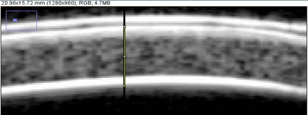
ImageJ measurement of central corneal thickness
This image processing program allows for measurement of clinically relevant structures such as the central corneal thickness, and also for measurement of structures that are not typically measured, such as the length of a ciliary process.

ImageJ measurement of ciliary processes
Some structural findings related to anterior segment anatomy (in particular the ciliary body) have unknown clinical significance. Unfortunately, current literature lacks prospective studies relating quantitative objective anterior segment structural data and long term outcomes. We hypothesize that the size and orientation of these under-studied anterior segment structures may be clinically meaningful. By use of a prospective analysis system, we hope to identify these significant structures.
Our measurement protocol has been put to the test in terms of reliability and reproducibility and has undergone multiple revisions so that clinicians worldwide can share a common protocol.